ZEISS Axiovert 5 FL Digital Digital Inverted Microscope - Product of Zeiss Taiwan Company
Order number
2252900220
Couldn't load pickup availability
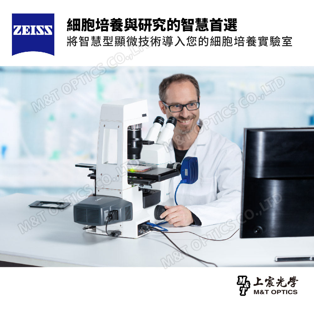
The smart choice for cell culture and research
ZEISS Axiovert 5 FL Digital is a digital smart inverted cell culture microscope that can obtain excellent imaging results in a short time. You just focus on visual observation, sample preparation, and executing your workflow. Axiovert 5 brings smart microscopy technology to your cell culture laboratory. Simply press the Snap button to save clear cell or tissue culture images. For transmitted light and multi-channel fluorescence imaging, this smart microscope automatically adjusts settings and parameters to fit the image. When shooting, the microscope automatically saves all image information such as scale and shooting parameters in the file.

Smart microscope that can work independently
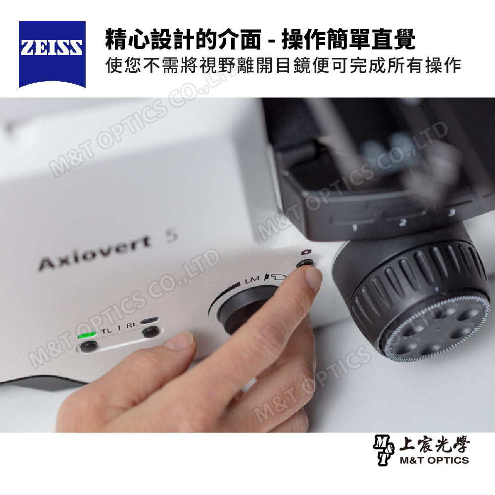
Axiovert 5 is equipped with a smart control box. You can use Axiovert 5 in standalone mode without any additional computer, as it is smartly integrated into the system. Control the microscope via the on-screen display (OSD) or use the ZEISS imaging application Labscope to take full advantage of the smart microscope. Camera setup, light source control and image enhancement are all automated.

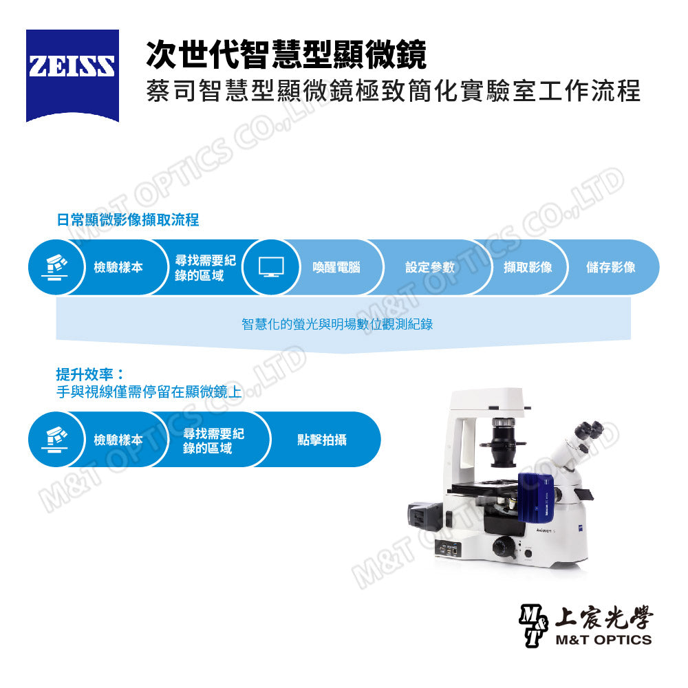
Next generation smart microscope - improve your work efficiency
Different from traditional laboratories, Zeiss smart microscopes extremely simplify laboratory workflow. With ZEISS Axiovert 5 FL Digital, you can automate your workflow and always focus on the sample. Once you find the target, press the button to record directly. Even without an external monitor, you can work smoothly and record observations. And, the microscope automatically determines the ideal settings for each channel. You get a superimposed multi-channel fluorescence image, with all associated image data automatically stored in your metadata. This process integrates perfectly with your established microscopy workflow, greatly increasing your efficiency.

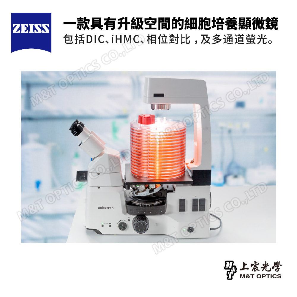
A cell culture microscope with room for upgrades
From routine cell culture to research, the ZEISS Axiovert 5 FL Digital fits seamlessly into your laboratory and workflow. Offers a wide range of possibilities for various applications. Paired with a compatible platform-top incubator, you can even use Axiovert 5 for long-term live cell imaging. If your work needs change, Axiovert 5 can adapt accordingly. With upgradable hardware and software, this smart microscope will be your reliable laboratory partner.

Scientific research grade ZEISS Axiocam Zeiss microscope imaging system
It leverages ZEISS' best microscopy image processing technology and perfectly combines the optical system of Zeiss microscopes with accurate colors, built-in brightness and flat-field calibration compensation functions, support for Zeiss' scientific-grade ZEN professional application software, and the extremely convenient Labscope mobile app. The device APP can perfectly control cameras and microscopes. Zeiss original factory will also provide an endless supply of high-value working modules and AI modules to keep your microscope system at the forefront of scientific research.

Supports multiple platforms and wireless transmission records
It can connect multiple computers and mobile devices via WiFi at the same time, and uses AI artificial intelligence to count cells and cell coverage. It can take pictures immediately with one click and generate the cell number and coverage. It can shoot continuously and quickly, and all image files are Complete information will be recorded, including objective lenses, camera systems, measurement scales, etc., and can be reviewed retrospectively at any time, enabling subsequent comparison and calculation of image data.

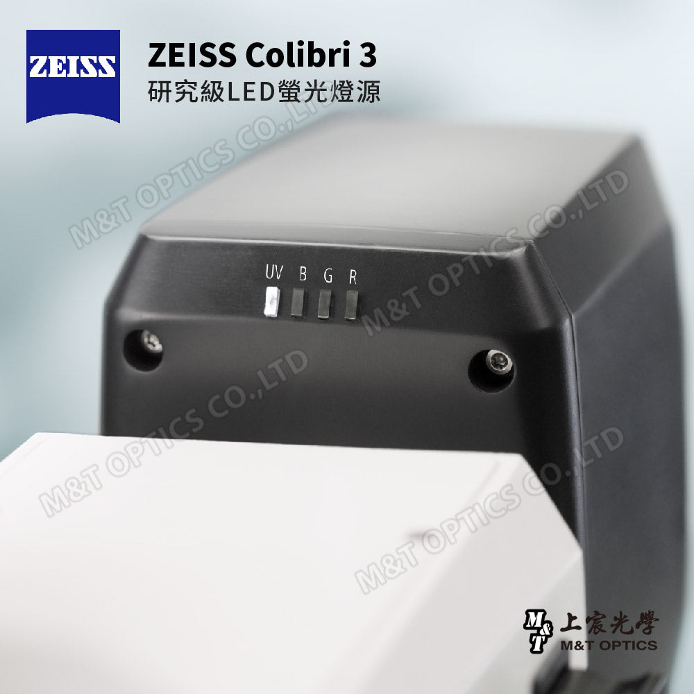
Colibri 3 - ZEISS Research Grade Fluorescent LED Lighting System
Colibri 3 makes it easy for you to obtain outstanding fluorescence images. You can easily switch between UV, blue, green and red excitation channels. Colibri 3 can stably output appropriate wavelength and intensity, gently excite fluorescent dyes and proteins, and does not harm the sample. To shoot, just select the corresponding channel and press the shutter.

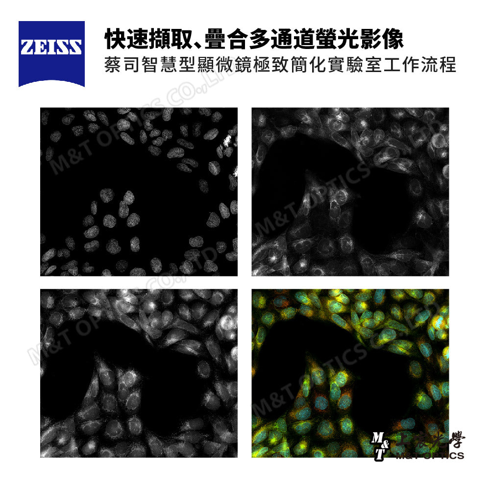
Capture five multi-channel fluorescence images with one click
Combining Axiovert 5 with the high-performance LED light source Colibri 3 and any ZEISS microscope camera gives you an excellent configuration for multi-channel fluorescence image recording. Easily switch between UV, blue, green and red excitation or transmitted light: simply select the corresponding channel and press the capture button. The system will automatically control the light source, adjust the exposure time, acquire images, switch channels, and then start again. That's it: you get superimposed multi-channel fluorescence images, including scale bars. Even without a computer. This condition is ideal for low fluorescence intensity applications such as transfection examinations.

Various digital recording functions and embedded camera system
ZEISS Axiovert 5 FL Digital can instantly output images under the microscope for fast photography, video recording and image processing. The operation interface has built-in scale, measurement and up to 15 kinds of labeling functions, which are fully prepared for the subsequent scientific analysis. You can also use a variety of different preset templates to quickly compile observation reports for easy sharing and recording, and even You don't need a computer, you just need to use an HDMI cable to connect the screen, and you can use your mouse to directly control the system built into the camcorder to take photos and videos, and record them on a flash drive.

Split windows for image comparison
The image file and the real-time microscope screen can be opened at the same time, and the two screens can be displayed at the same time to compare samples with each other and facilitate teaching demonstrations. When shooting, you can also capture the images in the split screen, output the observation report and annotate it.

AI artificial intelligence cell counting and coverage calculation
The AI artificial intelligence cell counting and real-time calculation and analysis of plate cell coverage, which have been trained by Zeiss global database, have achieved ultra-fast computing power with just one click. Not only is the accuracy extremely high, but the repeatability has also been verified by a large number of global professionals. Tested by users, it improves work efficiency and reduces the chance of personnel making mistakes when performing repetitive work.

Scan tissue samples easily
If your laboratory occasionally needs to scan tissue samples, Labscope's Fast Panorama module will be your first choice, converting your Axiovert 5 into a whole slide imaging system. By manually moving the microscope's sample stage, images of the sample are automatically stitched into a panoramic picture. This is the ideal solution when you need to scan whole slice images (WSI) from time to time. Whether you want to digitize an entire sample or just a portion of it at high resolution, Labscope's Fast Panorama module provides a simple and fast solution.

Virtual laser pen function
The virtual laser pen function can be activated, and the light point of the virtual laser pen can be projected onto a large screen or other connected device with a finger or cursor on the connected device, which is convenient for teaching and guidance, especially when explaining small structures and Interesting details for the occasion.

Tablet Micrograph-Augmented Reality Imaging Application
The auxiliary function of AR augmented reality technology is introduced, combined with the rear lens image of the tablet, allowing you to instantly compare the microscopic image being taken with a book or chart, or overlay it on a white paper to draw your own Observe the sample structure.

Observation case
HeLa Kyoto cells 63× LD Plan Neofluar dual-channel fluorescence image: nuclei are in blue and tubulin is in red

Observation case
HE-stained intestinal image under transmitted light, brightfield observation

ZEISS Axiovert 5 FL Digital parts description

Product Size

Product system diagram


Product specifications
| size | 503 × 363 × 505 ( L × W × H in mm ) |
| weight | 18.2kg |
| Integrated lighting system RGB-UV red (625 nm) | Excites Cy5, Alexa 631, TOTO-3 and similar dyes |
| Integrated lighting system RGB-UV green (565 nm) | Excites Cy3, TRITC, DsRed and similar dyes |
| Integrated lighting system RGB-UV blue (470 nm) | Excites eGFP, Fluo4, FITC and similar dyes |
| into lighting system RGB-UV UV (385 nm) | Excites DAPI, Alexa 405, Hoechst 33258 and similar dyes |
| transmitted illumination light | White 10W LED |
| focus | Coarse and fine focus with 13mm focuser and adjustable focus |
| condenser | LD condenser NA 0.4, includes phase slider for brightfield and phase contrast |
| photomicrography system | ZEISS Axiocam208 |
| pixel | 8.3M |
| Pixel size | 1.85 µm × 1.85 µm |
| Exposure time | 61 µs – 1 s |
| Sensitivity | 1x~22x |
| Photosensitive element size | 1/2.1"(8.1mm) |
| Signal output | HDMI, USB TYPE C 3.0, WiFi, WLAN |
| Software platform | Windows, Android, iOS |
| Warranty | One year for the whole machine |