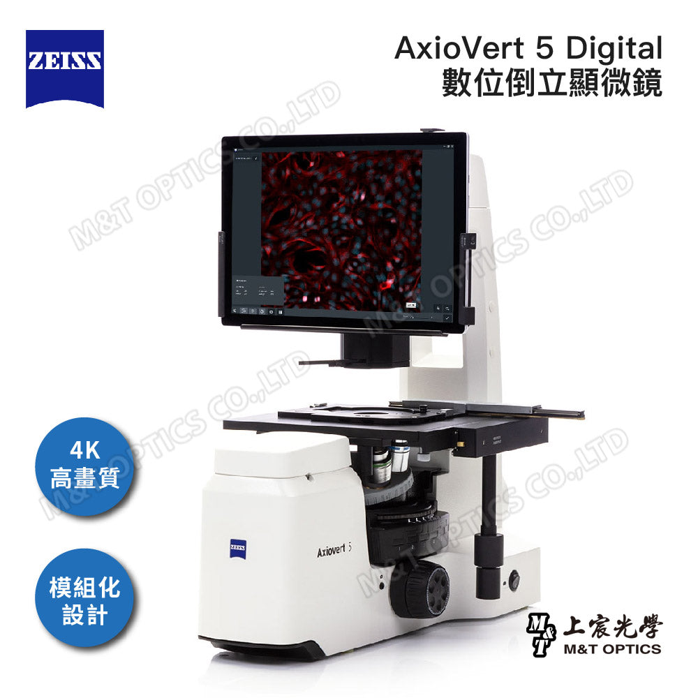
ZEISS Axiovert 5 Digital digital inverted microscope-original warranty company goods
Order number
2252900122
Couldn't load pickup availability

ZEISS AxioVert 5 Digital digital inverted microscope
Artificial intelligence (AI) is already helping our daily lives, from self-driving cars and home assistants to smartphones through facial recognition. Now, Axiovert 5 digital brings AI into your cell lab to simplify your daily work. It will make your process more efficient and produce clearer microscopic images.

simpler/smarter/modular
Explore creativity without being restricted or controlled by rules or traditions
Experience the full benefits of a modular microscope system. From scientific routine to basic research, phase contrast for multi-channel fluorescence imaging, even novice users are guaranteed to produce outstanding images with Axiovert 5 digital. All you have to do is turn on your system and focus on your sample. Don't worry about settings or adjustments - they're done automatically. Axiovert 5 digital will define new levels of reproducibility and data quality. You can always rely on the best performance of your instrument to produce publishable images.

Save time and let AI do the work for you
Saving time is easy with Axiovert 5 digital, which uses artificial intelligence to optimize and support daily workflows. Cell counting and cell fusion are automatically determined by off-the-shelf artificial intelligence modules. Now everyone can use AI in your lab: no training or prior knowledge required. Get instant results with just one click and they are absolutely replicable. Relax and enjoy watching the AI do the work for you. 
phase contrast cell lines

Analysis of cell lines using ZEISS Labscope

Made just for you
Axiovert 5 digital is the perfect choice for multi-user environments as proper system operation is supported by design. This all-in-one imaging system comes with an intuitive and simple operating concept. Triggered by pressing the Snap button.- Image acquisition of up to 5 channels, including multi-channel imaging
- AI cell counting and confluency workflow, ready for image analysis
- Video recording Axiovert 5 digital combines proven optical quality with ease of use

Make your cell experiments more reproducible
AI cell fusion and AI cell counting using the Zeiss Labscope module
If you work with cell cultures such as COS-7, HeLa, LoVo, or U2OS, you are probably familiar with tasks such as determining cell confluency and counting cells. These are critical values for your further decisions regarding cell proliferation, viability, adaptation to environmental conditions, harvesting cells, starting transfection, and preparing for experiments. Both cell confluency and count must be independent of cell shape, size, and type. Doing this manually can be time-consuming and laborious, resulting in an error-prone and subjective process.
It’s time to start making your experiments more reproducible, using pre-trained artificial intelligence to automatically analyze cell numbers and cell area covered. The ZEISS Labscope modules AI Cell Confluency and AI Cell Counting are ideally suited to your workflow. Check your cells as usual and simply take pictures as they move from one location to another in the cell culture container. Automatically analyze images and you'll receive intuitive, quantitative results instantly.

HeLa cell line, 20× objective | analyzed using Zeiss Labscope

Increase your efficiency with a smart microscope
Efficiency and quality are key in your laboratory, but acquiring multi-channel fluorescence images can be time-consuming. You need to: place the sample, focus on your area, switch to the computer, adjust the settings, then acquire the image, insert the scale bar, switch back to the microscope...and so on. Especially with manual microscopes, this process can be cumbersome. Imagine having a simple and painless way to acquire up to four fluorescence channels and one transmitted light channel, superimposed in a single image.With the Axiovert 5 digital smart microscope, the perfect settings for each channel are automatically determined. You get an overlaid multi-channel fluorescence image, with all relevant image data automatically stored in metadata. The program integrates perfectly with your established microscopy workflow, greatly increasing your efficiency.
ZEISS Labscope: Simple | Imaging | Application
The ZEISS Axiovert 5 digital is ready for use with ZEISS Labscope, the easy-to-use imaging software. Labscope has everything you need in the lab—from image acquisition, to clever built-in measurement capabilities to easy data sharing.

Get results quickly
Axiovert 5 digital offers an intuitive and clearly structured user interface. All important functions and parameters are directly visible or available with a single click. You can capture images, record videos, process imaging data, measure, annotate, and even generate reports including results.

Tailored to your application
Working in a busy laboratory, you need to be efficient. Whether acquiring large images of your entire slide in brightfield, multi-channel fluorescence imaging or observing your cells: Axiovert 5 digital is the best choice. Get fast results at the touch of a button.

Dedicated Labscope modules tailor-made to fit your application perfectly:
- Labscope AI Cell Fusion
- Labscope artificial intelligence cell counting
- Labscope quick panorama
- Labscope multi-channel

Transmitted light phase contrast is ideal for examining thin, unstained samples such as single cells.
Almost every cell biology experiment begins with cell culture. Whether primary cells or immortalized cell lines, the most important thing is health. and the normal behavior of cells before starting an experiment. This makes the contrast microscope the most important instrument for controlling your cell culture laboratory. Axiovert 5 digital is equipped with phase contrast to obtain high contrast images of cultured cells. You can observe and analyze your live cells without staining. With Axiovert 5 digital it is easy to implement the modules Labscope AI Cell Counting and AI Cell Confluency.
Multi-channel fluorescence: U2OS cells stained with NucBlue, CellMask green, MitoTracker red, phase contrast overlay.
Fluorophores and fluorescent proteins help characterize cellular structures and metabolic processes at the single-cell level microscopically and in situ. Without fluorescence microscopy, imaging differences between structures or even individual proteins would be unthinkable. Thanks to the Axiovert 5's integrated LED excitation unit digitally, you can get up to 4 fluorescence channels plus phase contrast at once. This can be used to obtain larger data sets with predefined automatic image acquisition lighting and camera settings.

In transmitted light field, you can quickly examine stained tissue sections.
Brightfield microscopy is the most common contrast microscopy technique. It is preferred for very thin tissue sections. Autologous specimens have little contrast and few structures are visible microscopically. Various staining methods are used to differentiate tissues. Here it is particularly important to record and reproduce structures with high contrast, while being able to distinguish even subtle color differences. Axiovert 5's digital built-in camera offers excellent resolution and high color fidelity. You can evaluate and annotate images directly using Labscope, even in live images.

Anti-theft clip prevents unauthorized disassembly of tablet | Tablet position can be independently adjusted for height and tilt

It is recommended to use a modern tablet to achieve the best experience | The tablet can be taken out when working

Install a light hood to block your sample from ambient light | With Axiovert 5 digital and Labscope, you can easily obtain high-quality fluorescence images.


Product specifications
| size | 503 × 363 × 505 ( L × W × H in mm ) |
|
weight
|
18.2kg |
| Integrated lighting system RGB-UV red (625 nm) | Excites Cy5, Alexa 631, TOTO-3 and similar dyes |
| Integrated lighting system RGB-UV green (565 nm) | Excites Cy3, TRITC, DsRed and similar dyes |
| Integrated lighting system RGB-UV blue (470 nm) | Excites eGFP, Fluo4, FITC and similar dyes |
| into lighting system RGB-UV UV (385 nm) | Excites DAPI, Alexa 405, Hoechst 33258 and similar dyes |
| transmitted illumination light | White 10W LED |
| focus | Coarse and fine focus with 13mm focuser and adjustable focus |
| condenser | LD condenser NA 0.4, includes phase slider for brightfield and phase contrast |
| Camera sensor type | SONY IMX264 Exmor Pregius Global Shutter CMOS |
|
Number of pixels
|
5.07 megapixels, 2464 (H) x 2056 (V) |
| Pixel size | 3.45 microns x 3.45 microns |
| Chip size | 8.5 mm x 7.1 mm, equivalent to 2/3'' (11.1 mm diagonal) |
|
Spectral range of black and white cameras
|
350nm to 1000nm |
| IR filter color camera | 400nm to 720nm |
|
Real time frame rate
|
25 frames per second at 1920x1080 pixels |